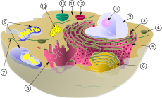
Centrosome
In cell biology, the centrosome is an organelle that is the main place where cell microtubules are organized. Also, it regulates the cell division cycle, the stages which lead up to one cell dividing in two.

(1) nucleolus
(2) nucleus
(3) ribosomes (little dots)
(4) vesicle
(5) rough endoplasmic reticulum (RER)
(6) Golgi apparatus
(7) Cytoskeleton
(8) smooth endoplasmic reticulum(SER)
(9) mitochondria
(10) vacuole
(11) cytoplasm
(12) lysosome
(13) centrioles within centrosome
The centrosome was discovered by Edouard Van Beneden in 1883,[1] and was described and named in 1888 by Theodor Boveri.[2]
The centrosome has apparently only evolved in animal cells.[3] Fungi and plants use other structures to organize their microtubules.[4] Although the centrosome has a key role in efficient mitosis in animal cells, it is not necessary.[5]
A centrosome is composed of two centrioles at right angles to each another. They are surrounded by a shapeless mass of protein.
Roles of the centrosome change
The centrosome is copied only once per cell cycle. Each daughter cell inherits one centrosome, containing two centrioles. The centrosome replicates during the interphase of the cell cycle. During the prophase of mitosis, the centrosomes migrate to opposite poles of the cell. The mitotic spindle then forms between the two centrosomes. Upon division, each daughter cell receives one centrosome.
Centrosomes are not needed for the mitosis to happen. When the centrosomes are irradiated by a laser, mitosis proceeds with a normal spindle. In the absence of the centrosome, the microtubules of the spindle are focused to form a bipolar spindle. Many cells can completely undergo interphase without centrosomes. It also helps in cell division.[6]
Although centrosomes are not needed for mitosis or the survival of the cell, they are needed for survival of the organism. Cells without centrosomes lack certain microtubules. With centrosomes the cell division is much more accurate and efficient. Some cell types arrest in the following cell cycle when centrosomes are absent, though this doesn't always happen.
References change
- ↑ Wunderlich V. 2002. JMM - past and present. Journal of Molecular Medicine 80 (9): 545–548. doi:10.1007/s00109-002-0374-y. PMID 12226736.
- ↑ Boveri, Theodor (1888). Zellen-Studien II: Die Befruchtung und Teilung des Eies von Ascaris megalocephala. Jena: Gustav Fischer Verlag.
- ↑ Bornens M.; Azimzadeh J. 2007. Origin and Evolution of the Centrosome. Advances in experimental medicine and biology 607: 119–129. doi:10.1007/978-0-387-74021-8_10. PMID 17977464.
- ↑ Schmit 2002. Acentrosomal microtubule nucleation in higher plants. International Review of Cytology 220: 257–89. doi:10.1016/S0074-7696(02)20008-X. PMID 12224551.
- ↑ Mahoney N.M.; Goshima G.; Douglass A.D.; Vale R.D. 2006. Making microtubules and mitotic spindles in cells without functional centrosomes. Current Biology 16 (6): 564–569. doi:10.1016/j.cub.2006.01.053. PMID 16546079.
- ↑ Rieder CI; Faruki S; Khodjakov A. 2001. The centrosome in vertebrates: more than a microtubule-organizing center. Trends in Cell Biology 11 (10): 413–9. doi:10.1016/S0962-8924(01)02085-2. ISSN 0962-8924. PMID 11567874.