File:Canine distemper pathology.jpg
Size of this preview: 466 × 599 pixels. Other resolutions: 187 × 240 pixels | 500 × 643 pixels.
Original file (500 × 643 pixels, file size: 68 KB, MIME type: image/jpeg)
| This is a file from the Wikimedia Commons. Information from its description page there is shown below. Commons is a freely licensed media file repository. You can help. |
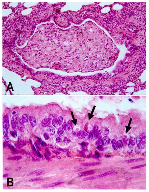
| Description | Lung lesions in an African wild dog with canine distemper. Hematoxylin and eosin staining. A. Bronchiole occluded by inflammatory cells and cell debris. B. Detail of A, showing multiple eosinophilic intracytoplasmic viral inclusions (arrows) in bronchiolar epithelium. |
| Source | Van de Bildt, Marco W.G.; Kuiken, Thijs; Visee, Aart M.; Lema, Sangito; Fitzjohn, Tony R.; Osterhaus, Albert D.M.E. Distemper Outbreak and Its Effect on African Wild Dog Conservation, 2002. Emerg Infect Dis [serial on the Internet]. 2002 Feb [cited 2007-06-18]. Available from https://www.cdc.gov/ncidod/eid/vol8no2/01-0314-G1.htm. |
| Author | |
| Permission (Reusing this file) |
Public Domain rationale |
This image is a work of the Centers for Disease Control and Prevention, part of the United States Department of Health and Human Services, taken or made as part of an employee's official duties. As a work of the U.S. federal government, the image is in the public domain.
|
File history
Click on a date/time to view the file as it appeared at that time.
| Date/Time | Thumbnail | Dimensions | User | Comment | |
|---|---|---|---|---|---|
| current | 22:09, 19 June 2007 | | 500 × 643 (68 KB) | Joelmills | {{Information |Description=Lung lesions in an African wild dog with canine distemper. Hematoxylin and eosin staining. A. Bronchiole occluded by inflammatory cells and cell debris. B. Detail of A, showing multiple eosinophilic intracytoplasmic viral inclus |
File usage
The following page uses this file:
Global file usage
The following other wikis use this file:
- Usage on ar.wikipedia.org
- Usage on de.wikipedia.org
- Usage on en.wikipedia.org
- Usage on es.wikipedia.org
- Usage on et.wikipedia.org
- Usage on eu.wikipedia.org
- Usage on fr.wikipedia.org
- Usage on ga.wikipedia.org
- Usage on he.wikipedia.org
- Usage on hu.wikipedia.org
- Usage on id.wikipedia.org
- Usage on ja.wikipedia.org
- Usage on ko.wikipedia.org
- Usage on uk.wikipedia.org
- Usage on www.wikidata.org
- Usage on zh.wikipedia.org