File:Fusiform face area face recognition.jpg
No higher resolution available.
Fusiform_face_area_face_recognition.jpg (475 × 503 pixels, file size: 95 KB, MIME type: image/jpeg)
| This is a file from the Wikimedia Commons. Information from its description page there is shown below. Commons is a freely licensed media file repository. You can help. |
Summary
| Description |
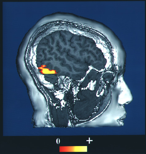
English: This is a computer-enhanced fMRI scan of a person who has been asked to look at faces. The image shows increased blood flow in the part of the visual cortex that recognizes faces.
日本語: fMRI。顔を見るように言われた人の脳内で、視覚皮質の顔情報を処理する部位で、血流増加が起きている、という画像。 |
| Date | upload to commons at 2009-10-27 |
| Source | https://www.nlm.nih.gov/hmd/emotions/frontiers.html (archive.org) |
| Author | NIH |
| Permission (Reusing this file) |
Public domain US government |
This image is a work of the National Institutes of Health, part of the United States Department of Health and Human Services, taken or made as part of an employee's official duties. As a work of the U.S. federal government, the image is in the public domain.
 |
||
| This file has been identified as being free of known restrictions under copyright law, including all related and neighboring rights. | ||
File history
Click on a date/time to view the file as it appeared at that time.
| Date/Time | Thumbnail | Dimensions | User | Comment | |
|---|---|---|---|---|---|
| current | 03:38, 27 October 2009 | | 475 × 503 (95 KB) | Was a bee | == {{int:filedesc}} == {{Information |Description= '''en:''' This is a computer-enhanced fMRI scan of a person who has been asked to look at faces. The image shows increased blood flow in the part of the visual cortex that recognizes faces. '''ja:'''fM |
File usage
The following page uses this file:
Global file usage
The following other wikis use this file:
- Usage on en.wikipedia.org
- Usage on es.wikipedia.org
- Usage on fa.wikipedia.org
- Usage on fr.wikipedia.org
- Usage on he.wikipedia.org
- Usage on hr.wikipedia.org
- Usage on hy.wikipedia.org
- Usage on it.wikipedia.org
- Usage on ja.wikipedia.org
- Usage on ko.wikipedia.org
- Usage on ml.wikipedia.org
- Usage on nl.wikipedia.org
- Usage on pl.wikipedia.org
- Usage on tr.wikipedia.org